Contribution of the Electron Channeling Contrast Imaging in polycrystal plasticity

Dr. Antoine GUITTON1,2

1Université de Lorraine – CNRS – Arts et Métiers – LEM3, Metz, France
2LabEx Damas – Université de Lorraine, Metz, France
antoine.guitton@univ-lorraine.fr
 www.antoine-guitton.fr


The full potential of ECCI for multiscale comparisons between experiments and simulations.

Although mechanics of materials is aged by more than one century, it still faces many conceptual challenges. One must relate two extreme scales: the sample scale (i.e. Macroscopic) and the scale of fundamental mech- anisms (i.e. Microscopic). In addition, statistics of observations are generally extremely low so that, on one hand the uncertainty is high and on the other hand the representativeness of mechanisms is very questionable.

The Transmission Electron Microscope (TEM) is one of the most well-known techniques for observing and characterizing dislocations in electron transparent thin foils (thickness of ≈ 100nm with a useful field of view of few μm) (1–4). Comprehensive dislocation studies at microscopic scale bring valuable information for extrapolating to the macroscopic mechanical response of materials and they can feed numerical advanced multiscale crystal plasticity models (4). However, fundamental questions on the representativeness of observed phenomenon must be raised, when extrapolating discussions to a millimeter-size specimen.

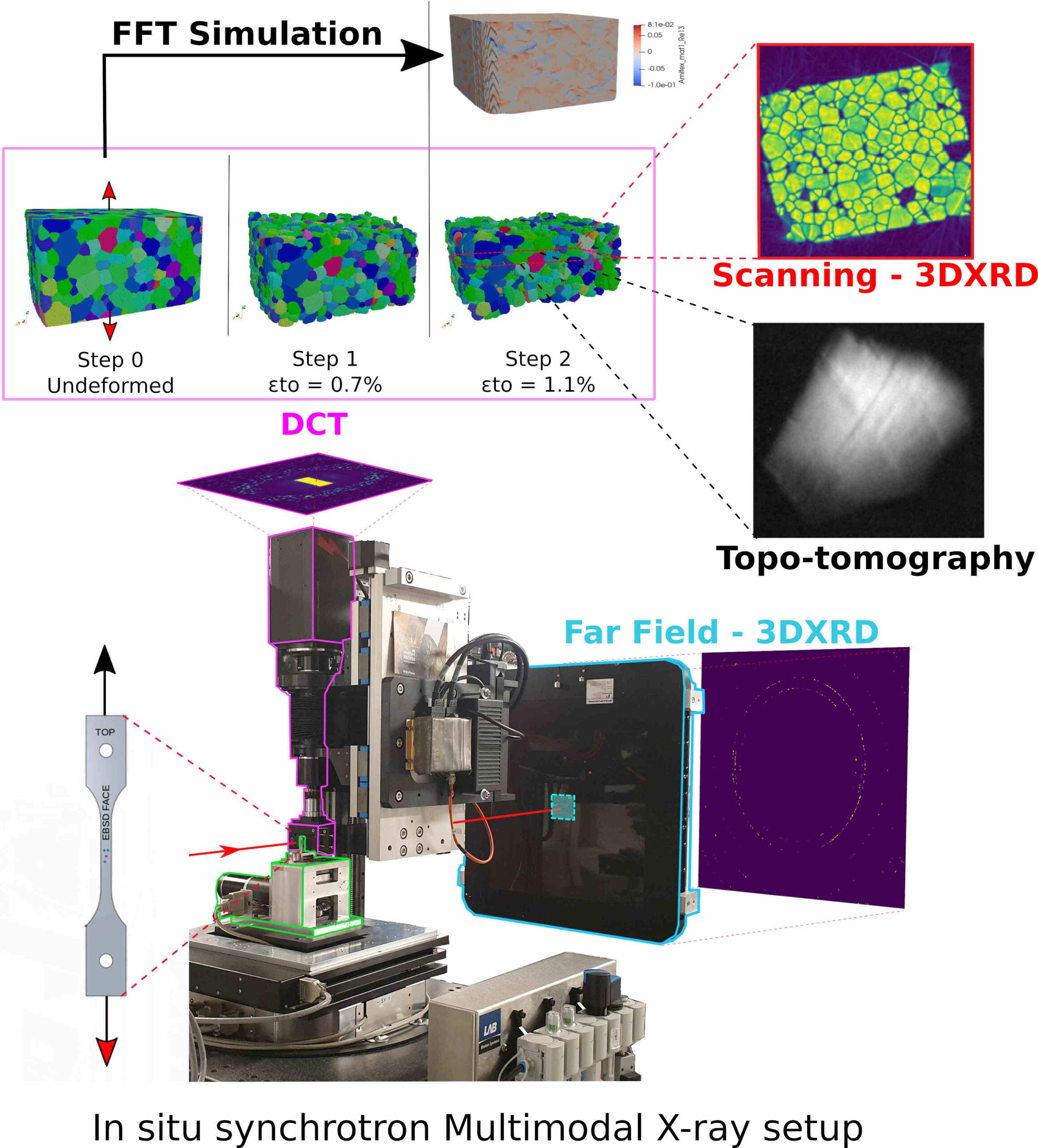
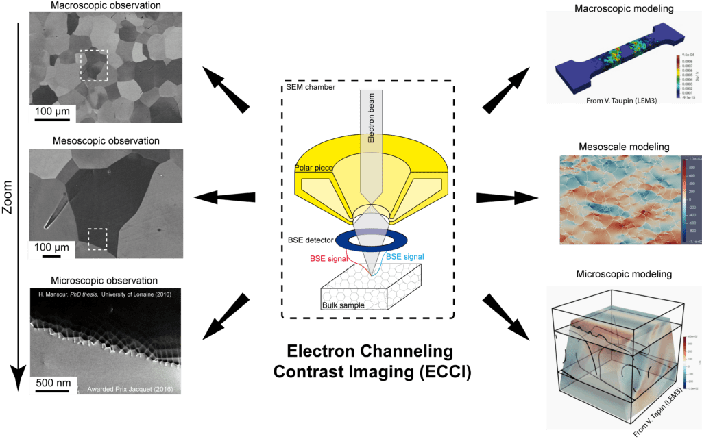
In this framework, we have successfully combined mechanical testing (nanoindentation and in-situ tensile tests) of bulk specimens with a dislocation-scale characterization technique: Accurate Electron Channeling Contrast Imaging (A-ECCI) (5). A-ECCI is a non-destructive procedure offering the ability to provide, inside a Scanning Electron Microscope (SEM), TEM-like diffraction contrast imaging of sub-surface defects (at a depth of about 100 nm) on centimetric bulk specimen with still unsurpassed resolutions (6).

First, physics of defect contrasts and experimental procedures will be presented (5; 7; 8). Second, the full potentiality of A-ECCI for following the evolution of deformation microstructures will be detailed (9–11). Finally, statistical comparisons between crystal plasticity models and experiences will be highlighted in the framework of a first step towards feature engineering (12–14).

Thursday 27 May 2021 14h00

videoconference at the following link: https://rdv.onera.fr/seminaireLEM

References

[1]  G.P. Bei, A. Guitton, A. Joulain, V. Brunet, S. Dubois, L. Thilly, and C. Tromas. Pressure-enforced plasticity in MAX phases: from single grain to polycrystal investigation. Philosophical Magazine, 93(15):1784–1801, may 2013, doi: 10.1080/14786435.2012.755272, hal-hal-01501851.

[2]  A. Guitton, A. Joulain, L. Thilly, and C. Tromas. Dislocation analysis of Ti2AlN deformed at room temperature under confining pressure. Philosophical Magazine, 92(36):4536–4546, dec 2012, doi: 10.1080/14786435.2012.715250, hal-03041046.

[3]  A. Guitton, A. Joulain, L. Thilly, and C. Tromas. Evidence of dislocation cross-slip in MAX phase deformed at high temperature. Scientific Reports, 4(1):6358, may 2015, doi: https://doi.org/10.1038/srep06358, hal-01503720.

[4]  K. Gouriet, P. Carrez, P. Cordier, A. Guitton, A. Joulain, L. Thilly, and C. Tromas. Dislocation modelling in Ti2AlN MAX phase based on the Peierls–Nabarro model. Philosophical Magazine, 95(23):2539–2552, aug 2015, doi: 10.1080/14786435.2015.1066938, hal-01515323.

[5]  H. Kriaa, A. Guitton, and N. Maloufi. Fundamental and experimental aspects of diffraction for characterizing dislocations by electron channeling contrast imaging in scanning electron microscope. Scientific Reports, 7(1):9742, aug 2017, hal-02392256.

[6]  J. Guyon, H. Mansour, N. Gey, M.A. Crimp, S. Chalal, and N. Maloufi. Sub-micron resolution selected area electron channeling patterns. Ultramicroscopy, 149:34–44, feb 2015, doi: 10.1016/j.ultramic.2014.11.004, hal-01514962.

[7]  H. Kriaa, A. Guitton, and N. Maloufi. Modeling dislocation contrasts obtained by Accurate-Electron Channeling Contrast Imaging for characterizing deformation mechanisms in bulk materials. Materials, 12(10):1587, may 2019, doi: 10.3390/ma12101587, hal-02392249.

[8]  H. Kriaa, A. Guitton, and N. Maloufi. Modelling Electron Channeling Contrast intensity of stacking fault and twin boundary using crystal thickness effect. Materials, 14(7):1696, mar 2021, doi: 10.3390/ma14071696, hal-03118996.

[9]  A. Guitton, H. Kriaa, E. Bouzy, J. Guyon, and N. Maloufi. A dislocation-scale characterization of the evolution of deformation microstructures around nanoindentation imprints in a TiAl alloy. Materials, 11(2):305, feb 2018, doi: 10.3390/ma11020305, hal- 02392252.

[10]  M. Ben Haj Slama, N. Maloufi, J. Guyon, S. Bahi, L. Weiss, and A. Guitton. In situ macroscopic tensile testing in SEM and Electron Channeling Contrast Imaging: pencil glide evidenced in a bulk β-Ti21S polycrystal. Materials, 12(15):2479, aug 2019, doi: 10.3390/ma12152479, hal-02392248.

[11]  F. Habiyaremye, A. Guitton, F. Sch ̈afer, F. Scholz, M. Schneider, J. Frenzel, G. Laplanche, and N. Maloufi. Plasticity induced by nanoindentation in CrCoNi medium-entropy alloy studied by accurate electron channeling contrast imaging revealing dislocation-low angle grain boundary interactions. Accepted in Materials Science and Engineering: A, 2021, hal-03118990.

[12]  M. Ben Haj Slama, V. Taupin, N. Maloufi, K. Venkatraman, A.D. Rollett, R.A. Lebensohn, S. Berbenni, B. Beausir, and A. Guitton. Electron channeling contrast imaging characterization and crystal plasticity modelling of dislocation activity in Ti21S BCC material. Materialia, page 100996, mar, doi: 10.1016/j.mtla.2020.100996, hal-03094460.

[13]  K. Venkatraman, M. Ben Haj Slama, V. Taupin, N. Maloufi, and A. Guitton. Tuning critical resolved shear stress ratios for BCC- Titanium Ti21S via an automated data analysis approach. 2021, hal-03119000.

[14]  F. Habiyaremye, A. Guitton, X. Lei, T. Richeton, S. Berbenni, G. Laplanche, and N. Maloufi. Influence of the local dislocation density and configuration on the first pop-in load during instrumented nanoindentation. 2021.

Characterisation of shear bands and plasticity in model glasses at the atomic scale

First synthesised in the 60’s, the metallic glasses are a very promising class of material thanks to their very high yield strength. Yet, these materials are also very brittle due to the formation of persistent shear bands which concentrate plastic deformation.

In this thesis, we perform atomistic simulations with a simple two-dimensional binary Lennard-Jones model glass. To link plasticity and the material structure, we use a novel structural indicator, the local yield stress. 

Through this measure, the material average local yield stress is shown to increase as the degree of relaxation increases. We also find the existence of a unique post-yield shear threshold distribution, independent on the initial state of the material.

By the mean of an elementary model, the origin of the Bauschinger effect in amorphous solids (a plasticity-induced  asymmetry of the mechanical behaviour) is found to arise from the inversion of the low yield barriers population anisotropy during the unloading.

Then, by considering systems of different sizes and degrees of relaxation the persistence of plasticity, and thus the formation of shear-bands, is shown to mostly depend on the degree of relaxation of the system.

Finally, in well relaxed glasses, a correlation between the location of the shear band and the initial soft regions is shown. As further loading is applied on the material, a diffusive broadening of the shear band is observed.

Modeling of platinum-based nano-alloys: Co-Pt, emblematic system of the order, and Pt-Ag, hybrid system between order and demixtion.

Due to the strong correlation between chemical order and physical properties, nanoalloys with a tendency to order are particularly interesting in the field of catalysis, magnetism, or optics. By reducing the size of the system, i.e. from a solid alloy to a nanoalloy, many questions arise: Is the chemical order preserved? What is the morphology of nanoparticles? What is the composition and chemical order on the surface? What is the evolution of properties with size? This presentation is devoted to the study of two systems, both similar and different in their behavior: Co-Pt, a system emblematic of the chemical order, and Pt-Ag, a hybrid system presenting both a chemical order and a tendency to demix, as well as a strong tendency to segregation. In order to answer these various questions, we adopt a semi-empirical approach through an N-body potential, allowing atomic relaxations, in the approximation of the second moment of state density (SMA), coupled with Monte Carlo simulations in different ensembles. The SMA potential is adjusted, in order to reproduce the volume and surface properties, on calculations derived from the theory of density functional theory (DFT) or on experimental data. In a first step, the volume phase diagram of the two systems is determined by the model and compared to the experiment. Then the low index surfaces (111), (100) and (110) are studied in order to verify the segregation inversion observed for the Co-Pt system, where Pt segregates weakly on the dense surfaces (111) and (100) but where we observe a pure Co plane on the surface (110). On the contrary, the Pt-Ag system shows strong Ag segregation on surfaces (111) and (100). In a second step, aggregates of truncated octahedral morphology of different sizes (ranging from 1000 to 10000 atoms) will be analyzed in terms of chemical composition on the different unequal sites (top, edge, facets (100) and (111) and core) and then compared to the reference systems (surfaces, volume) over the whole concentration range. For the Co-Pt system, we observe ordered structures similar to those of the volume for the core and similar to those of the surfaces for the facets. The impact of the two-dimensional phase (√3 × √3)R30◦ specific to the surface, is all the more important on the chemical order at the core as the nanoparticle is small. For the Pt-Ag system, we observe an important segregation of Ag at the surface, as well as a Pt enrichment at the subsurface, and the stabilization of the L11 ordered phase at the core. This structure can appear in a single variant or by adopting all possible variants, leading to an onion peel structure.


CPFEM simulations of grain size effect in FCC polycrystals: a new approach based on surface GND density

A multiscale modeling methodology involving discrete dislocation dynamics (DDD) and crystal plasticity finite element method (CPFEM) is used to study the physical origin and to simulate the grain size effect in FCC polycrystalline plasticity. This model is based on the dislocation density storage–recovery framework, expanded on the scale of slip systems. DDD simulations are used to establish a constitutive law incorporating the main dislocation mechanisms controlling strain hardening in monotonically deformed FCC polycrystals. This is achieved by calculating key quantities controlling the accumulation of the forest dislocation density within the grains and the polarized dislocation density at the grain boundaries during plastic deformation. The model is then integrated into the CPFEM at the polycrystalline aggregate scale to compute short- and long-range internal stresses within the grains. These simulations quantitatively reproduce the deformation curves of FCC polycrystals as a function of grain size. Because of its predictive ability to reproduce the Hall-Petch law, the proposed framework has a great potential for further applications.

Speaker: Maoyuan Jiang

Date and Location: Monday 09/03/20 14h00, LEM meeting room (E2.01.20), Châtillon.

Orientation imaging at the onset of plastic deformation


Diffraction Contrast Tomography (DCT) is a near-field X-ray diffraction technique for the inspection of ductile materials at the micron scale. It has traditionally been used for the study of undeformed polycrystalline materials with grain sizes of a few tenths of microns. It uses a box-sized monochromatic X-ray beam, which allows it to scan large regions of millimeter sized sample (with up to thousands of grains) in a relatively short time.
Recent work has introduced sub-grain orientation reconstruction (6D-DCT), which has made DCT a viable tool for the reconstruction of slightly deformed materials.
Topo-tomography (TT) is also a near-field X-ray diffraction technique, which, on the other hand, allows to focus on a single grain with a high-resolution detector and to obtain sub-micron level shape information.
In this talk, we will first present how the data is acquired and reconstructed in modern DCT and TT acquisitions. Then, we will present their 6D and 5D extensions (respectively) for the reconstruction of sub-grain level orientation information. Finally, we will discuss future applications, including the combined use of DCT and TT data in a single 6D reconstruction for the investigation of slip bands formation at the onset of deformation.

Speaker: Dr Nicola Viganò

Date and Location: Friday 21/02/20, 14h00 LEM meeting room (E2.01.20), Châtillon.

Coupling Bragg Coherent Diffraction Imaging (BCDI) and Molecular Dynamics to investigate nanostructure

Fig. 1 (top) Experimental reconstruction of the u111 displacement field on a 250 nm Pt NP (bottom)  u111 displacement field obtained by energy minimization of a simulated Pt NP (right) εxx, εyy and  εzz components of the strain tensor derived from the simulation

Physical properties at small length scale deviate strongly from the bulk counterpart, typically below the micrometer. For instance, mechanical strength increases with reducing size, large residual strain due to processing are present in nanostructures. Thus a better understanding of the physical properties in relationship with the microstructure is needed for nanoscale materials. Because of its good spatial resolution (~ 10 nm) and excellent sensitivity to atomic displacements and local strain [1,2], Bragg coherent diffraction imaging (BCDI) has emerged in the past two decades as a powerful tool to probe the structure and local displacement field inside nanoscale objects [3]. When combined with in situ mechanical loading, BCDI is particularly relevant for the study of defect nucleations in isolated nanoparticles [4] or to investigate intragranular deformation mechanisms in polycrystalline thin films [5].

Nowadays, the length scales that are accessible by BCDI and that can be simulated by Molecular Dynamics (MD) simulation are almost converging. The coupling between the two methods is therefore particularly relevant and allows to get a detailed picture of the deformation mechanisms in nanostructures at the atomic scale. This coupled approach has been used to study the surface relaxation of metallic nanoparticles (Au, Pt). An excellent quantitative agreement is obtained between the component of the displacement field measured experimentally and calculated by energy minimization (Molecular Statics) (Fig. 1). With this approach, the measurement of only one Bragg reflection is required to derive the 3D displacement field and the six independent components of the strain tensor from the simulation [6]. The two techniques can also be combined to  identify defect structures nucleated during in situ  mechanical loading [4,5] and to interpret the evolution of the strain field in nanoparticle catalysts during gas reaction [7,8]..

[1] Watari, M. et al. Nature Materials 10, 862–866 (2011).

[2] Labat, S. et al. ACS Nano 9, 9210–9216 (2015).

[3] Robinson, I. & Harder, R. Nat Mater 8, 291–298 (2009).

[4] Dupraz, M. et al. Nano Lett. 17(11) (2017).

[5] Cherukara, M. et al. Nat. Comm. (2018).

[6] Dupraz et al.  to be submitted (2019)

[7] Kim, D. et al. Nat. Comm. 9, 3422 (2018).

[8] Dupraz, M. et al. in preparation

Speaker: Dr. Maxime Dupraz

Date and Location: Monday 25/11/19, 14h00 LEM meeting room (E2.01.20), Châtillon.

2018, web site created by HA & RG.